Oral Pathology and Oncology
Comprehensive diagnosis and management of oral lesions with emphasis on early intervention.


Understanding Oral Pathology and Oncology
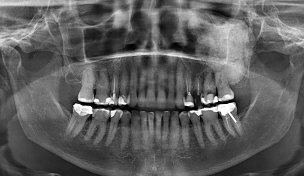
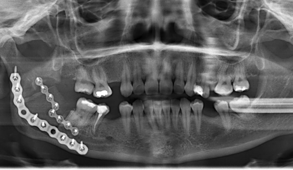
Oral pathology and oncology in maxillofacial and oral surgery focus on diagnosing and managing abnormal tissue growths in the mouth and jaw, including benign cysts, tumors, and oral cancer. These specialised fields address concerning changes such as red or white patches, persistent sores, lumps, or difficulties with chewing and swallowing.
Early identification and treatment are critical to prevent progression to more serious disease. The primary goals are to remove abnormal growths, confirm diagnosis through biopsy, and in cases of malignancy, prevent further spread while restoring oral function and appearance.
Oral Pathology and Oncology Indications
You may require evaluation and treatment if you have:
- Persistent red (erythroplakia) or white (leukoplakia) patches in your mouth
- Non-healing ulcers or sores that bleed easily
- Lumps, thickenings, or abnormal tissue growth in the oral cavity
- Chronic sore throat, hoarseness, or difficulty swallowing
- Suspicious lesions identified during dental check-ups or self-examinations
Any persistent change in your oral tissues that lasts more than two weeks should be evaluated by a healthcare professional. Early detection significantly improves treatment outcomes, particularly for oral cancer.
Oral Pathology and Oncology Approach
Oral pathology and oncology treatment works through a systematic approach to diagnosis and management:
- Diagnostic Precision: Thorough examination and biopsy confirm the exact nature of abnormal tissues
-
- Targeted Removal: Surgical techniques remove pathological tissues while preserving healthy structures
-
- 3D Planning: Advanced imaging and modeling ensure precise surgical approaches
-
- Multidisciplinary Integration: Collaboration with specialists in radiation and medical oncology when needed
-
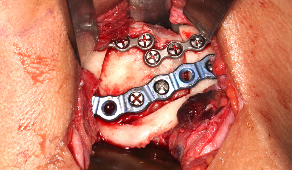
- Functional Reconstruction: Custom solutions restore oral function and appearance after tissue removal
The approach is tailored to the specific condition, with treatment intensity proportional to the severity and type of pathology diagnosed.
Oral Pathology and Oncology Protocol
1. Comprehensive Assessment and Diagnosis
- Detailed clinical examination of suspicious areas
- Imaging studies (X-rays, CT scans, MRI) to determine the extent of the lesion
- Biopsy to confirm the nature of the tissue (benign, precancerous, or malignant)
2. Treatment Planning
- Development of a customised treatment approach based on diagnosis
- For complex cases, creation of 3D-printed models to guide surgical planning
- Multidisciplinary consultation for cases requiring combined therapy
3. Surgical Intervention
For Benign Conditions
- Excision of cysts or benign tumors
- Curettage (scraping) of affected bone areas
- Marsupialization of large cysts when appropriate
For Precancerous Lesions
- Complete removal with careful margin evaluation
- Close follow-up to monitor for recurrence
For Malignant Tumors
- Wide excision with appropriate margins
- Lymph node evaluation when indicated
- Immediate reconstruction planning using CAD-designed implants
4. Adjunctive Therapies (for malignant cases)
- Coordination with oncologists for chemotherapy
- Radiation therapy planning when indicated
- Targeted biological therapies in specific cases
5. Reconstruction and Rehabilitation
- Restoration of oral function using grafts or implants
- Speech and swallowing therapy when needed
- Dental rehabilitation to restore chewing function
Oral Pathology and Oncology Recovery
Recovery varies significantly based on the type and extent of treatment:
- Minor Biopsies and Small Lesion Removal:
- Healing typically occurs within 1-2 weeks
- Minimal disruption to daily activities
- Benign Tumor or Cyst Removal:
- Initial recovery period of 1-3 weeks
- Complete healing within 4-8 weeks depending on size and location
- Major Resections and Reconstructions:
- Hospital stay of 5-14 days depending on complexity
- Initial recovery period of 4-6 weeks
- Adaptation to reconstruction and return to function over 3-6 months
- Combined Surgery and Radiation/Chemotherapy:
- Radiation typically begins 4-6 weeks after surgery
- Complete treatment course may extend 3-6 months
- Full recovery may take 6-12 months
- Long-term Follow-up:
- Regular monitoring for recurrence
- Ongoing care of reconstructed areas
- For malignant conditions, surveillance continues for several years
- Minor Biopsies and Small Lesion Removal:
Dr Fakir will provide personalised recovery guidelines based on your specific diagnosis and treatment plan. Throughout your recovery, you’ll receive comprehensive support to ensure optimal healing and restoration of oral function.